A Closer Look: Evaluating Mohs Surgery’s Role in the Treatment of Invasive Melanoma of the Head and Neck

2024
In this Perspectives on the Science piece, we reflect on the implications of the recently conducted multicenter study on the treatment of invasive head and neck melanoma using Mohs micrographic surgery.
Authors
Affiliations

Ching, Lauren

Georgetown University School of Medicine

Strong, Jennifer

National Institutes of Health

Lee, Truelian

Harvard Medical School

Kaufman, Howard L.

Massachusetts General Hospital

Harvard Medical School

Emerick, Kevin S.

Mass Eye and Ear

Harvard Medical School

Kim, Emily Y.

Brigham and Women’s Hospital

Patel, Vishal A.

GW School of Medicine & Health Sciences

Brownell, Isaac

National Institutes of Health

Singh, Kritika

National Institutes of Health

Neel, Victor A.

Massachusetts General Hospital

Harvard Medical School

Miller, David Michael

Massachusetts General Hospital

Harvard Medical School

Gupta, Sameer

Mass Eye and Ear

Harvard Medical School

Published

January 24, 2024

Doi

Featured Article


Brandon T. Beal, Jeremy Udkoff, Leora Aizman, Jeremy Etzkorn, John A. Zitelli, Christopher J. Miller, Thuzar M. Shin, Joseph F. Sobanko, David G. Brodland, Outcomes of invasive melanoma of the head and neck treated with Mohs micrographic surgery – A multicenter study, Journal of the American Academy of Dermatology, Volume 89, Issue 3, 2023, Pages 544-550, ISSN 0190-9622, https://doi.org/10.1016/j.jaad.2022.12.038.


Introduction

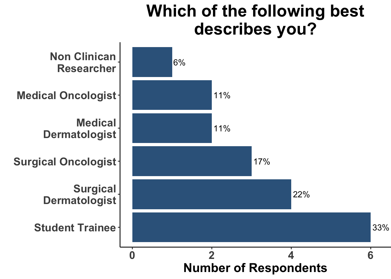
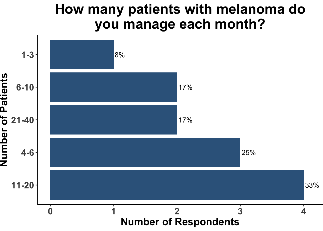
On November 6th, 2023 the multi-institutional Society of Cutaneous Oncology (SoCO) Journal Club (JC) reviewed the Journal of American Academy of Dermatology article “Outcomes of invasive melanoma of the head and neck treated with Mohs micrographic surgery – A multicenter study”1. Participants included clinicians and investigators from Massachusetts General Hospital, Massachusetts Eye and Ear, the National Institutes of Health, the Dana-Farber Cancer Institute, George Washington University School of Medicine, Georgetown University School of Medicine and the University of Pittsburgh (Figure 1), with a range of experience managing melanoma (Figure 2). This Perspectives on the Science article reflects the authors’ views after the Journal Club. Please note that it does not represent the views of other SoCO members or affiliated institutions. In this article, we provide a summary of the article as well as discuss its strengths and limitations.

Figure 1: Attendees at the November 6th SoCO Journal Club were surveyed as to their professional role.

Background for the Study

The management of invasive melanoma on the head and neck presents significant challenges. These challenges include unique surgical locations, positive excision margins, local recurrence, a roughly 10% risk each for upstaging and a tenfold increased likelihood of a flap or graft reconstruction, often referred to as the rule of 10s.2

Presently, the National Comprehensive Cancer Network (NCCN) guidelines for invasive melanoma stem from findings across six randomized control trials (RCTs) involving a total of 4,206 cases of melanoma with a mere 27 (0.6%) of these of these being located on the head or neck (Table 1).39 Thus, the current body of consistent clinical data guiding the selection of surgical management or margins for invasive melanoma in the head and neck (H&N) region is limited.

Studies Evaluating Surgical Margins in Melanoma
Study Overview Participants (n) H&N Melanoma Local Recurrence Nodal Recurrence Distant Metastasis Disease Specific Survival
Veronesi 19911 Limited Excision: 1 cm vs. Wide Excision: 2 cm Pimary: ≤2.0 mm thick 612 0 (0%) Limited Excision: 4 (1.31%) Wide Excision: 0 Limited Excision: 21 (6.89%) Wide Excision: 24 (7.81%) Total: 45 (7.35%) Limited Excision: 17 (5.57%) Wide Excision: 14 (4.56%) Narrow excision: 81.6% Wide Excision: 84.4% (8-year DFS rates)
Cohn-Cedermark et al., 20002 Limited Excision: 2 cm vs. Wide Excision: 5 cm Primary: >0.8-≤ 2.0 mm thick 989 0 (0%) Limited Excision: 3 (0.6%) Wide Excision: 5 (1%) Limited Excision: 70 (15%) Wide Excision: 61 (12%) Limited Excision: 71 (15%) Wide Excision: 70 (15%) N/A
Balch et al., 20013 Limited Excision: 2 cm vs. Wide Excision: 4 cm Primary: 1-4 mm thick 740 64 (8.65%) Group A: Limited Excision: 0.4% Wide Excision: 0.6% Group B: Limited Excision: 9.93% N/A N/A Limited Excision: 70% Wide Excision: 77% (10-year DFS rates)
Khayat et al., 20024 Limited Excision: 2 cm vs. Wide Excision: 5 cm Primary: <2.1 mm thick 337 16 (4.75%) Limited Excision: 1 (0.62%) Wide Excision: 4 (2.4%) Limited Excision: 13 (8.1%) Wide Excision: 11 (6.7%) Limited Excision: 4 (2.5%) Wide Excision: 10 (6.1%) Limited Excision: 85% Wide Excision: 83% (10-year DFS rates)
Thomas et al., 20045 Limited Excision: 1 cm vs. Wide Excision: 3 cm Primary: ≥2 mm thick 900 0 (0%) Limited Excision: 15 (3.31%) Wide Excision: 13 (2.9%) Limited Excision: 135 (29.8%) Wide Excision: 118 (26.4%) Limited Excision: 38 (8.39%) Wide Excision: 30 (6.71%) Limited Excision: 48.6% Wide Excision: 43.6% (8-year DFS)
Gillgren et al., 20116 Limited Excision: 2 cm vs. Wide Excision: 4 cm Primary: >2 mm thick 936 0 (0%) Limited Excision: 20 (4.3%) Wide Excision: 9 (1.91%) Limited Excision: 100 (21.5%) Wide Excision: 114 (24.2%) Limited Excision: 38 (8.17%) Wide Excision:54 (11.5%) Limited Excision: 56% of cohort Wide Excision: 56% of cohort (5-year DFS rates)
Utjes et al., 20197 Limited Excision: 2 cm vs. Wide Excision: 4 cm Primary: >2 mm thick 936 0 (0%) N/A N/A N/A HR: 0.95 (0.78-1.16)
References: 1 Veronesi (1991) 2 Cohn-Cedermark, Rutqvist, Andersson, Breivald, Ingvar, Johansson, J�nsson, Krysander, Lindholm, and Ringborg (2000) 3 Balch, Soong, Smith, Ross, Urist, Karakousis, Temple, Mihm, Barnhill, Jewell, Wanebo, and Desmond (2001) 4 Khayat, Rixe, Martin, Soubrane, Banzet, Bazex, Lauret, Vérola, Auclerc, Harper, and Banzet (2003) 5 Thomas, Newton-Bishop, A'Hern, Coombes, Timmons, Evans, Cook, Theaker, Fallowfield, O'Neill, Ruka, and Bliss (2004) 6 Gillgren, Drzewiecki, Niin, Gullestad, Hellborg, Månsson-Brahme, Ingvar, and Ringborg (2011) 7 Utjés, Malmstedt, Teras, Drzewiecki, Gullestad, Ingvar, Eriksson, and Gillgren (2019)

Table 1: Data from clinical studies investigating the role of surgical margins in melanoma. Studies conducted in the first-line setting are highlighted in red. Abbreviations: CITN, cancer immunotherapy trials network; CTLA4, cytotoxic T-lymphoctye associated protein 4; IST, investigator-sponsored trial; NA, not available; PD-1, programmed cell death protein 1; PD-L1, programmed cell death ligand 1.

The traditional approach of treating H&N melanomas using conventional excision (CE) can be challenging because the anatomic sites may not easily accommodate wide excisions margins of 1-2 cm. In addition, there is data to suggest that CE may result in increased rates of positive margins, local recurrences, and disease specific deaths compared to CE for invasive trunk and extremity melanoma. Mohs micrographic surgery (MMS), a technique long used in the management of non-melanoma skin cancer, has increasingly been incorporated in the treatment of in situ, as well as invasive, melanoma (Tables 2-3).1,1036 Some reports have demonstrated that MMS can be a more comprehensive method for assessing peripheral and deep surgical margins during melanoma treatment.34,37,38 A retrospective study involving 36 cases of melanoma in situ treated with the method of serial cross-sectioning used in these standard evaluations at intervals of 1, 2, 4, and 10 mm could only detect positive margins 58%, 37%, 19%, and 7% of the time, respectively.39 Such data provide rationale for adopting complete histological margin control to reduce the risk of recurrence significantly. Staged excision allows for comprehensive histological margin control, aiming to reduce the risk of recurrence more effectively. One of the limitations of staged excisions is that procedures may extend over days to weeks.

Mohs for Melanoma In Situ
Study Overview Participants (n/N) H&N Melanoma Local Recurrence Nodal Recurrence Distant Metastasis Disease Specific Survival
Robinson 19941 Surgical margins for lentigo maligna 16/16 16 (100%) 1 NR 0 NR
Cohen et al., 19942 MMS for lentigo maligna 26/45 Unclear (41/45 LM and LMM) 0 NR 0 NR
Zitelli et al., 19973 MMS for treatment of primary cutaneous melanoma 176/535 106 (57.6%) 1 (0.5%) NR 1 99% (5-year)
Cohen et al. 19984,5 MMS on patients with lentigo maligna 54/62 NR 0 NR 0 NR
Bricca et al., 20056 MMS for cutaneous head and neck MIS 331/625 331 (100%) NR NR 1 99.2% (5-year)
Bhardwaj et al., 20067 MMS for lentigo maligna 158/200 132/158 (84%) 0 NR 0 NR
Temple et al., 20068 MMS for lentigo maligna 119/202 tumors Unclear (185/196 tumors) 0 NR Unclear (2 total) NR
Walling et al., 20079 Staged excision vs. MMS for lentigo maligna and lentigo maligna melanoma 16/57 (14/18 LM) 16 (89%) 6 NR 0 NR
Bene et al., 200810 MIS on the head and neck 167/167 (116 lentigo maligna, 51 MIS) 145 (87%) 2 NR 0 NR
Shumaker et al., 200911 MMS for periocular MIS 18/29 18 (100%) 3 NR NR NR
Hilari et al., 201112 MMS vs. conventional surgery for lentigo maligna on the head Unclear/57 (41 or 45/62 tumors) 41 or 45 (100%) 2 NR NR NR
Kunishige et al., 201213 Surgical margins for MIS 1072/1072 634 (56%) 3 NR 5 99.5% (5-year), 99.2% (10-year)
Newman et al., 201314 MMS for lentigo maligna using immunostaining 193/260 155 (80%) 3 NR 1 NR
Hou et al., 201515 Outcomes of MMS and wide excision for primary lentigo maligna Unclear/407 (154/423 tumors) 148 (96%) 3 NR NR 96% (5-year), 94% (10-year), 94% (15-year)
Etzkorn et al., 201516 MMS for MIS Unclear (436/597 tumors) 347/436 (80%) 2 NR NR NR
Felton et al., 201617 MIS on the head and neck 343/343 343 (100%) 1 NR 0 NR
Valentin-Nogueras et al., 201618,5 MMS in treatment of invasive melanoma and MIS 863/1982 NR 4 NR 0 100% (5-year)
Dika et al., 201619 MMS vs. video-assisted MMS vs. surgical excision for head and neck lentigo maligna 29/54 29 (100%) 1 NR 0 NR
Stigall et al., 201620 MMS for MIS of trunk and proximal extremities 882/882 tumors 0 (0%) 1 1 1 NR
Nosrati et al., 201721 Comparison of MMS and wide local excision for MIS 277/662 245 (88%) 5 NR 1 92% (5-year)
Foxton et al., 201822 MMS for lentigo maligna 62/62 55 (89%) 0 NR NR NR
Kunishige et al., 201923 Surgical margins for MIS vs. lentigo maligna 2335/2335 (1506 lentigo maligna, 829 MIS) 1362 (58%) 6 NR 0 NR
Demer et al., 20195 MMS vs. wide local excision for head and neck MIS 220/388 NR 2 NR 1 NR
References: 1 Robinson (1994) 2 Cohen, McCall, Hodge, Freedman, Callen, and Zax (1994) 3 Zitelli, Brown, and Hanusa (1997) 4 Cohen, McCall, and Zax (1998) 5 Demer, Vance, Cheraghi, Reich, and Lee (2019) 6 Bricca, Brodland, Ren, and Zitelli (2005) 7 Bhardwaj, Tope, and Lee (2006) 8 Temple and Arlette (2006) 9 Walling, Scupham, Bean, and Ceilley (2007) 10 Bene, Healy, and Coldiron (2008) 11 Shumaker, Kelley, Swann, and Greenway H. T. (2009) 12 Hilari, Llorca, Traves, Villanueva, Serra-Guillén, Requena, Llombart, Sanmartín, Guillén, and Nagore (2012) 13 Kunishige, Brodland, and Zitelli (2012) 14 Newman, Beal, Schram, and Lee (2013) 15 Hou, Reed, Knudson, Mirzoyev, Lohse, Frohm, Brewer, Otley, and Roenigk (2015) 16 Etzkorn, Sobanko, Elenitsas, Newman, Goldbach, Shin, and Miller (2015) 17 Felton, Taylor, and Srivastava (2016) 18 Valentín-Nogueras, Brodland, Zitelli, González-Sepúlveda, and Nazario (2016) 19 Dika, Fanti, Christman, Piraccini, Misciali, Vaccari, and Patrizi (2015) 20 Stigall, Brodland, and Zitelli (2016) 21 Nosrati, Berliner, Goel, McGuire, Morhenn, de Souza, Yeniay, Singh, Lee, Nakamura, Wu, Griffin, Grimes, Linos, Chren, Grekin, and Wei (2017) 22 Foxton, Elliott, and Litterick (2018) 23 Kunishige, Doan, Brodland, and Citelli (2019)

Table 2. Data from clinical studies investigating outcomes of MMS for MIS. Abbreviations: MMS, Mohs micrographic surgery; IM, invasive melanoma; LM, lentigo maligna; LMM, lentigo maligna melanoma; MIS, melanoma in situ; WLE, wide local excision; NR, not reported

Mohs for Invasive Melanoma
Study Overview IM Participants (n/N) H&N Melanoma Local Recurrence Nodal Recurrence Distant Metastasis Disease Specific Survival
Cohen et al., 19941 Rush permanent sections for LM & LMM 19/45 NR 0% (0/19) 0% (0/19) 0% (0/19) 100%
Zitelli et al, 19972 MMS for primary cutaneous melanomas 369/535 125 (33.9%) 1.4% (5/369) NR NR 93% (5-year)
Cohen et al., 19983 Follow up to Cohen et al, 1994 19/38 NR 0% (0/19) 0% (0/19) 5.3% (1/19) 100%
Clayton et al, 20004 Rush permanent sections for melanocytic neoplasms 25/119 24/25 (96%) 0% (0/25) NR NR NR
Bhardwaj et al, 2006 MMS with Mel5 for LM vs LMM 42/200 37 (88.1%) 0% (0/42) 0% (0/42) 0% (0/42) 100%
Temple & Arlette, 20065 MMS for LM vs LMM 69/199 NR 0% (0/69) NR NR NR
Moehrle et al, 20066 MMS vs WLE for LMM 292/292 238 (81.5%) 6.4 (10/156) for WLE vs 0.7% (1/136) for MMS 7.1% (11/156) for WLE vs 0.7% (1/136) for MMS 9.6% (15/156) for WLE vs 0.7% (1/136) for MMS 88.97% for WLE vs 100% MMS
Shumaker et al, 20097 Rush permanent sections for periocular melanoma 11/29 11 (100%) 18.2% (2/11) NR 27.3% (3/11) 72.7% (5-year)
Newman et al., 20138 MMS with Mel5 for LM & LMM 67/260 52 (20%) 0% (0/67) 0% (0/67) 0% (0/67) 100% (13-year)
Chin-Lenn et al., 20139 MMS vs WLE for IM 151/151 151 (100%) 7.7% (7/91) WLE vs 5.2% (3/60) MMS (p=.69) NR 7.2% WLE vs 8.1% MMS (p= .49) 82.8% WLE vs 92.4% MMS (p = .59)
Etzkorn et al., 201510 MMS with MART-1 for MIS and IM 161/561 128 (79.50%) 0% (0/161) NR NR NR
Valentin-Nogueras et al., 201611 MMS with MART1 for MIS and IM 556/2114 NR 1.3% (7/556) NR NR 96.42 (5-year)
Demer et al., 201912 MMS vs WLE for H&N melanoma 160/389 160 (100%) 6.7% (6/89) for WLE vs 0% (0/71) for MMS NR 21% (19/89) WLE vs 5.6% (4/71) MMS 86.5% WLE vs 98.6% MMS
Cheraghlou et al., 201913 MMS vs WME for early stage IM 3234/70319 1294 (40.0%) NR NR NR 79.2% (10-year), HR 0.86
Degesys et al., 201914 MMS with MART1 for IM 123/123 70 (56.9%) 1.63% (2/123) NR 0% (0/123) 100%
Heath et al., 202015 En face permanent sectioning for MIS vs IM 128/657 113 (88.28%) 0% (0/128) NR 1.6% (2/125) 99% (5-year)
Shin et al., 202116 MMS with MART-1 for MIS and IM 507/1790 NR 0.19% (1/507) NR NR NR
Burnett et al., 202117 MMS for IM of trunk and proximal extremities 1416/1416 0 (0%) 0.14% (2/1416) 1.91% (27/1416) 2.0% (28/1416) 96.3%
Vieira et al., 202218 MMS without immunostaining 43/234 25 (58.14%) 6.98% (3/43) NR NR NR
Beal et al., 202319 MMS with MART1 for H&N IM 785/785 785 (100%) 0.51% (4/785) 1.0% (8/785) 1.1% (9/785) 93.4% (10-year)
References: 1 Cohen, McCall, Hodge, Freedman, Callen, and Zax (1994) 2 Zitelli, Brown, and Hanusa (1997) 3 Cohen, McCall, and Zax (1998) 4 Clayton, Leshin, Hitchcock, Marks, and White (2000) 5 Temple and Arlette (2006) 6 Moehrle, Dietz, Garbe, and Breuninger (2006) 7 Shumaker, Kelley, Swann, and Greenway H. T. (2009) 8 Newman, Beal, Schram, and Lee (2013) 9 Chin-Lenn, Murynka, McKinnon, and Arlette (2013) 10 Etzkorn, Sobanko, Elenitsas, Newman, Goldbach, Shin, and Miller (2015) 11 Valentín-Nogueras, Brodland, Zitelli, González-Sepúlveda, and Nazario (2016) 12 Demer, Vance, Cheraghi, Reich, and Lee (2019) 13 Cheraghlou, Christensen, Agogo, and Girardi (2019) 14 Degesys, Powell, Hsia, and Merritt (2019) 15 Heath, Woody, Leitenberger, Latour, and Bar (2020) 16 Shin, Nugent, Aizman, Weiner, O'Malley, Sobanko, Etzkorn, Chu, Elenitsas, Giordano, Higgins II, and Miller (2021) 17 Burnett, Brodland, and Zitelli (2021) 18 Vieira, Jennings, Renzi M. A., Lawrence, and Decker (2022) 19 Beal, Udkoff, Aizman, Etzkorn, Zitelli, Miller, Shin, Sobanko, and Brodland (2023)

Table 3. Data from clinical studies investigating outcomes of MMS for IM. Studies with available outcome data for invasive melanoma were included. Abbreviations: MMS, Mohs micrographic surgery; IM, invasive melanoma; LM, lentigo maligna; LMM, lentigo maligna melanoma; WLE, wide local excision; NR, not reported; MIS, melanoma in situ; WME, wide margin excision.

In contrast, MMS may offer complete margin assessment during melanoma treatment without delay in subsequent reconstruction.34,37,3941 The initial use of MMS – one form of complete margin control – faced challenges given concern, by some, in accurately identifying melanocytes in frozen sections. However, the utilization of immunohistochemical (IHC) stains, especially Melanoma Antigen Recognized by T cells (MART-1), to aid in frozen section analysis has made the identification of melanoma reliably possible.37,38 Frozen sections using MART-1 have been shown to provide equivalent detection of melanoma as MART-1 stained permanent sections.42 A recent survey showed that 91% to 97.8% of Mohs surgeons trained in the last two decades are now using MART-1 as their exclusive choice for IHC staining during MMS on melanoma.37

These findings highlight the potential value of integrating MMS and IHC techniques in the treatment of melanoma, particularly in the head and neck region.

Study Design

This retrospective study, conducted at Zitelli & Brodland, P.C. and the University of Pennsylvania across 12 years, reviewed 785 cases of invasive melanomas in the head and neck region, treated with MMS using MART-1 immunohistochemical stains. Eligible participants were adults with biopsy-confirmed primary invasive melanoma on the head and neck, absent of metastatic disease on presentation. Excluded from the study were melanoma in situ, tumors on the trunk or extremities, previously treated cases, and participants presenting with nodal or distant metastasis.

Study Results

The study encompassed the treatment of 785 melanoma cases with variances in thickness between 0.3mm and 8.5mm through MMS. Following treatment, recurrence details were analyzed which revealed that local recurrence was observed in 0.51% (approximately 4 out of 785 cases), nodal recurrence was seen in 1.0% (approximately 8 out of 785 cases), and distant recurrence in 1.1% (approximately 9 out of 785 cases). When delving into the recurrence rates according to tumor stages T1 to T4, the study indicated that local recurrence stood at 0.16% for T1 stage (1 out of 636 cases), 1.18% for T2 stage (1 out of 85 cases), 2.22% for T3 stage (1 out of 45 cases), and 5.26% for T4 stage (1 out of 19 cases) (Figure 3). The survival rates after five and ten years were also promising, with disease-specific survival calculated at 96.8% (with a 95% confidence interval between 95.0% and 98.5%) and 93.4% (with a 95% confidence interval between 88.5% and 98.3%) respectively.

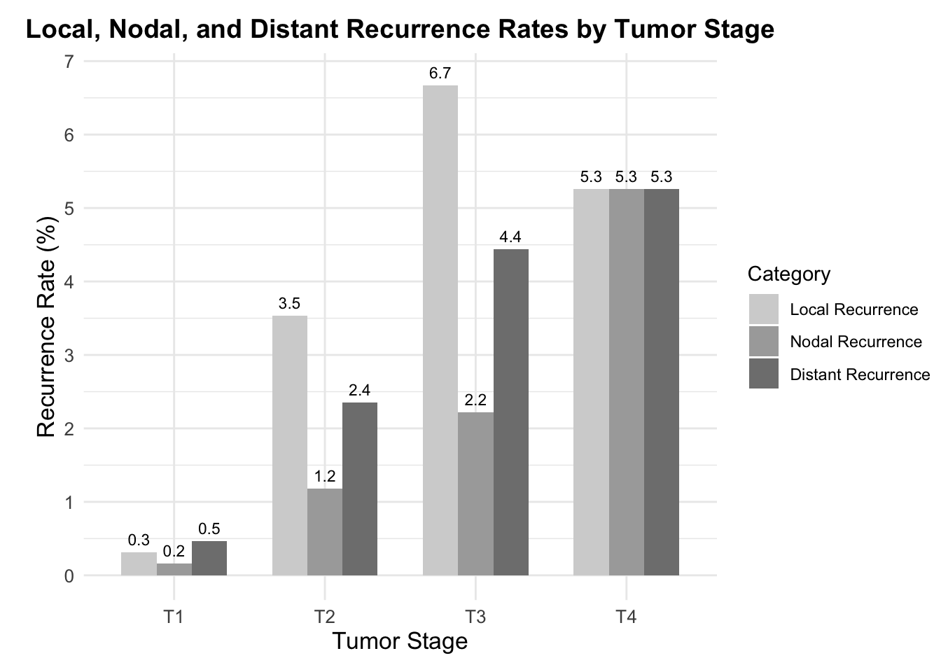
Figure 3. Comparative Recurrence Rates for Different Tumor Stages. The chart showcases Local Recurrence (LR), Nodal Recurrence (NR), and Distant Recurrence (DR) rates for tumor stages T1, T2, T3, and T4. Each recurrence type is represented by a different shade of gray, allowing for a clear comparison of recurrence rates across each tumor stage.

Discussion

The potential for same-day complete surgical margin analysis in head and neck invasive melanomas presents potential benefits compared to the conventional excisional method. These advantages include (1) a more complete resection of malignant cells due to more thorough margin assessment; and (2) achieving negative margins prior to reconstruction without delay. The use of a staged excision protocol may indeed achieve the former, but not necessarily the latter. Whether the application of MMS provides equal or superior results to conventional methods is yet to be elucidated with prospective data.

Beal et al. 2023 provides retrospective data from two institutions exploring the use of MMS for invasive melanoma. Strengths of this study include a sizable cohort of 785 cases treated with MMS for invasive melanomas on the head and neck over a 12-year period. Two distinct clinical sites using similar surgical techniques were included without discernible differences in terms of local recurrence (LR), nodal recurrence (NR), and distal recurrence (DR). This suggests that the results are not specific to a single practice. Given that the majority of patients (81%) had a Breslow depth of 1 mm or less, the low overall LR, NR, and DR noted in the study for invasive melanoma may be most confidently inferred for early invasive disease.

The limitations of the study include a variable follow-up method, with some individuals followed up through phone calls while others were considered based on their last physical examination recorded in their medical records. The study also lacked a matched control group for a clear comparison, or even a historical control population. There was a shortage of detailed reporting on certain clinical characteristics such as the number of surgical stages required for the complete clearance of the tumor.

Patients in the study eligible for sentinel lymph node biopsies (SLNB) based on the NCCN guidelines were offered testing; however, this data was not tracked, which would have been helpful in understanding the reported recurrence rates. Also not included was the technical challenges of performing SLNB in conjunction with MMS; understanding how this was or was not incorporated would also be helpful in understanding the generalizability of this approach. SLNB data has demonstrated relevance in providing an accurate prognostication as well as potential improvements in disease management in the context of a shifting therapeutic landscape, which might influence recurrence rates.41,43 The study included patients up until 2017 during a time when the management of metastatic melanoma was advancing due to the emergence of approval for immune checkpoint inhibitors (ICI) and BRAF and MEK inhibitors. The U.S FDA, in 2011, approved the use of the anti–CTLA-4 antibody ipilimumab for patients with stage IV melanoma, and later in 2014 approved anti–PD-1 antibodies nivolumab and pembrolizumab for second line treatment. These therapies have shifted the survival rate from less than 10% to 30% to 50% over the past decade.40,44 Additionally, in 2015, the NCCN recommended, and the FDA approved the use of PD-1 ICI as the first line therapy for patients with stage IV melanoma. More recently, immunotherapy and targeted therapy has also been approved for adjuvant therapy, including patients with Stage IIB-IV melanoma. Understanding the influence of these novel therapies on patient survival rates necessitates further research.

Another limitation is that the study did not directly compare MMS to CE or staged excision. A comparative analysis would have been useful to assess the relative effectiveness of the two strategies and would have reinforced their support of MMS for invasive melanoma on the head and neck rather than comparing against historical data. The retrospective, non-randomized design of the study was also a subject of discussion, leading to an agreement on the need for future prospective studies.

Inputs were sought from the experienced, multi-disciplinary SoCO group regarding their preferred treatment modality based on hypothetical clinical scenarios. In consideration of a case involving a 71-year-old male patient with a 1 cm wide, 1.1 mm thick melanoma on the vertex scalp, a quarter of the participants expressed a preference for MMS as their treatment method while the remaining 75% supported the conventional excision technique. Another case, involving the same measurements of melanoma but located on the right forearm of the male patient, saw unanimous consensus for conventional excision as the treatment method (Figures 4 and 5).

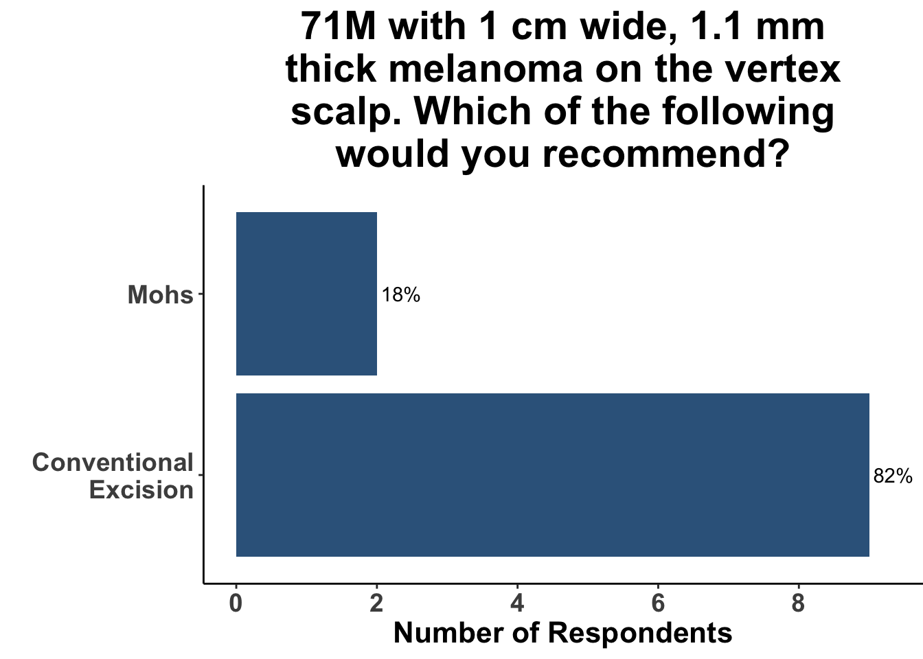
Figure 4: Journal club attendees were queried as to how they would manage a patient with Head and Neck Melanoma. The answer choice “I am not a clinician” was omitted (n = 6).

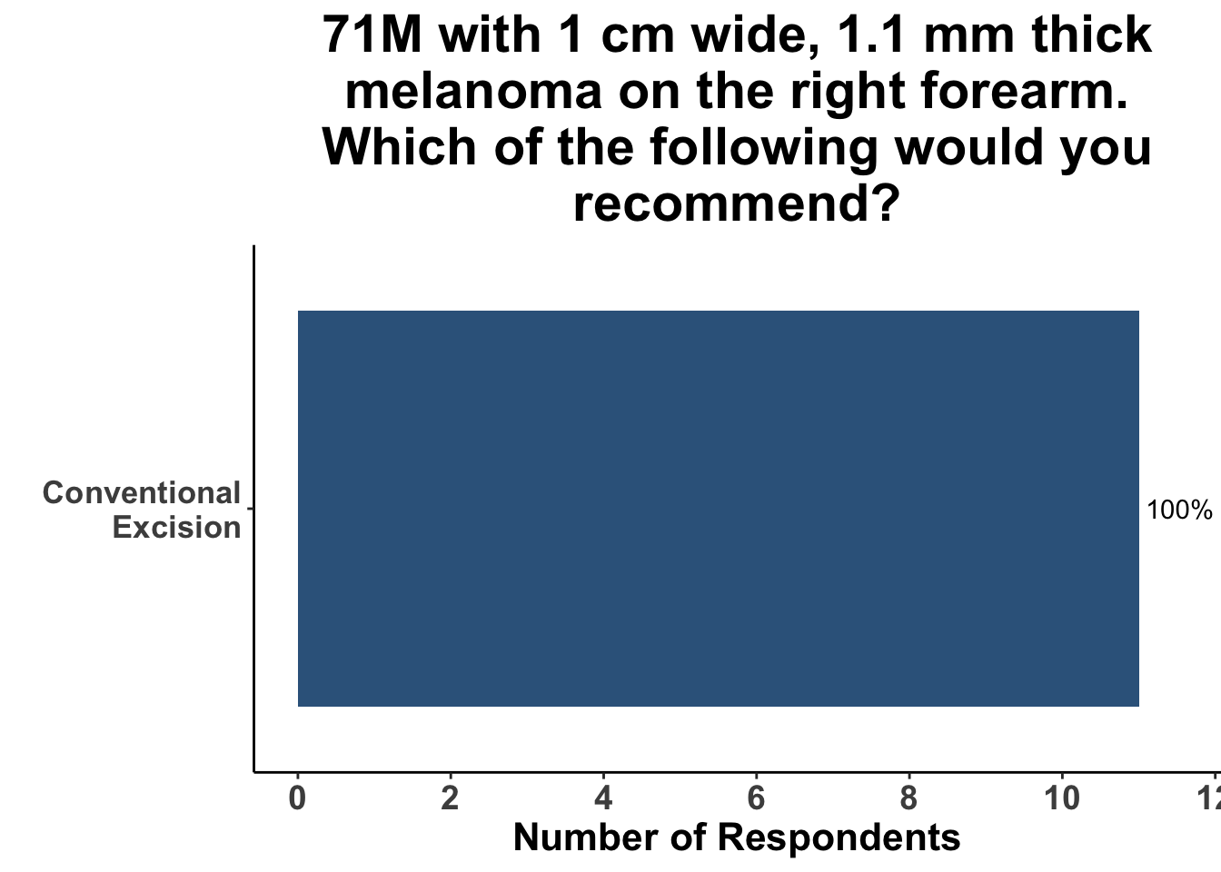
Figure 5: Journal club attendees were queried as to how they would manage a patient with melanoma on the forearm. The answer choice “I am not a clinician” was omitted (n = 6).

After discussion of the article, 17% of respondents felt the study provided adequate evidence for a shift towards MMS for the treatment of invasive head and neck melanomas (Figure 6). In terms of the utility of MMS over conventional excision, one-fifth of the clinicians anticipated potential changes in their medical practice (Figure 7). A majority of the participants (91%) felt that while MMS for invasive head and neck melanoma may show promise, challenges remain in terms of incorporating MMS into their own practice (Figure 8). This suggests the opportunity for prospective randomized trials or even matched case-control studies comparing surgical interventions.

Given that the SoCO group practitioners form a diverse group with various experiences managing melanoma, the survey data collected gives insight into the acceptance and potential deterrents to integrating MMS into mainstream practice.

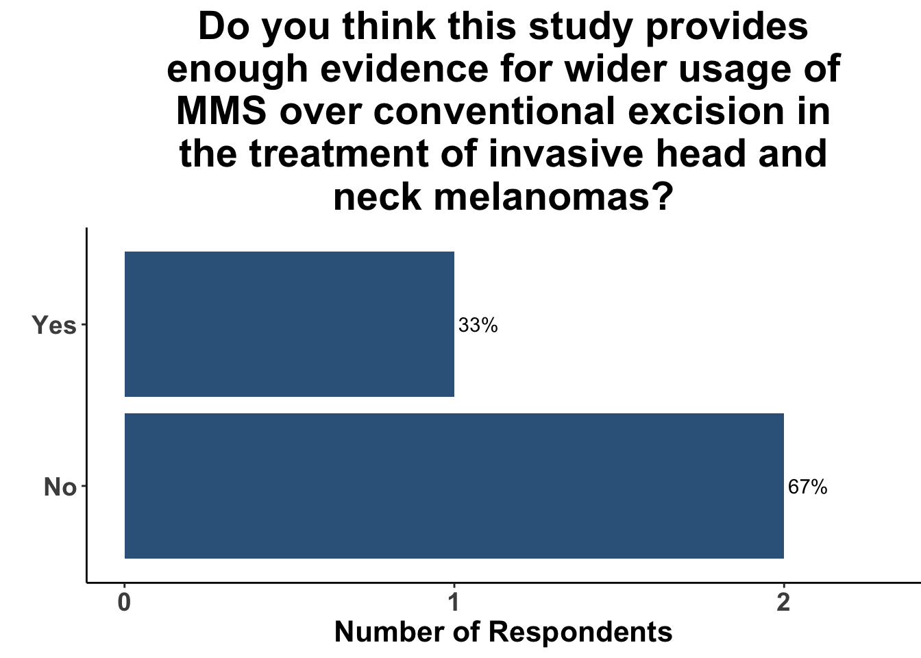
Figure 6: Journal club attendees were queried as to whether they thought the study by Beal et al. provided enough information to implement Mohs micrographic surgery over conventional excision. Abbreviations: MMS, Mohs micrographic surgery.

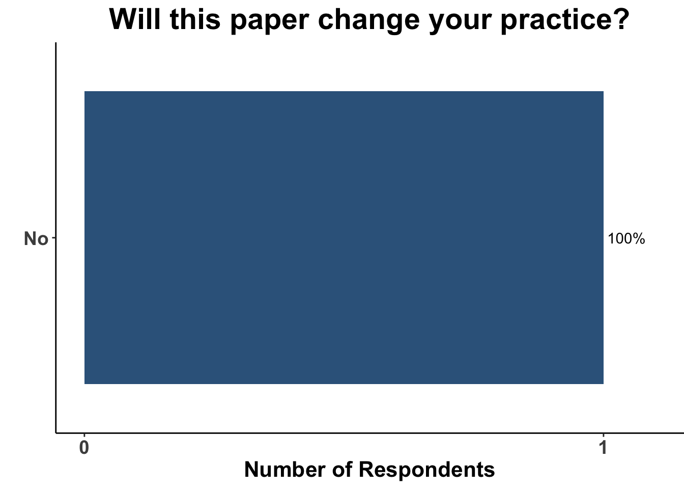
Figure 7: Journal club attendees were queried as to whether or not the study by Beal et al. would change their practice. The answer choice “I do make recommendations for melanoma patients” was omitted (n = 7) from the graph.

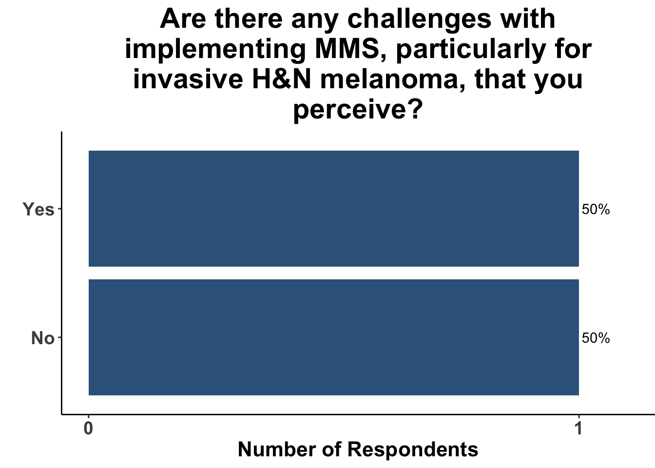
Figure 8: Journal club attendees were queried as to whether they perceived any challenges with implementing MMS, particularly for invasive H&N melanoma, into their practice. Abbreviations: H&N, head and neck; MMS, Mohs micrographic surgery.

Materials and Methods

This Perspectives on the Science piece was published using Quarto®. The survey was conducted using REDCap®.45 The figures depicting the survey data were created using R (version 4.0.0) and the tidyverse suite of packages,46 including ggplot2.47 The image on the “Perspectives on the Science” page was created by the authors (DMM) using the rosemary package.48

Bibliography

1.
Beal, B. T. et al. Outcomes of invasive melanoma of the head and neck treated with Mohs micrographic surgery A multicenter study. Journal of the American Academy of Dermatology 89, 544–550 (2023).
2.
3.
Veronesi, U. Narrow Excision (1-cm Margin). Archives of Surgery 126, 438 (1991).
4.
5.
6.
7.
8.
Thomas, J. M. et al. Excision Margins in High-Risk Malignant Melanoma. New England Journal of Medicine 350, 757–766 (2004).
9.
10.
Robinson, J. K. Margin control for lentigo maligna. J Am Acad Dermatol 31, 79–85 (1994).
11.
Zitelli, J. A., Brown, C. & Hanusa, B. H. Mohs micrographic surgery for the treatment of primary cutaneous melanoma. J Am Acad Dermatol 37, 236–245 (1997).
12.
Cohen, L. M., McCall, M. W. & Zax, R. H. Mohs micrographic surgery for lentigo maligna and lentigo maligna melanoma. A follow-up study. Dermatol Surg 24, 673–677 (1998).
13.
Bricca, G. M., Brodland, D. G., Ren, D. & Zitelli, J. A. Cutaneous head and neck melanoma treated with mohs micrographic surgery. J Am Acad Dermatol 37, 236–245 (2005).
14.
Walling, H. W., Scupham, R. K., Bean, A. K. & Ceilley, R. I. Staged excision versus Mohs micrographic surgery for lentigo maligna and lentigo maligna melanoma. Journal of the American Academy of Dermatology 57, 659–664 (2007).
15.
Bene, N. I., Healy, C. & Coldiron, B. M. Mohs micrographic surgery is accurate 95.1. Dermatol Surg 34, 660–664 (2008).
16.
Shumaker, P. R., Kelley, B., Swann, M. H. & Greenway, Jr., H. T. Modified mohs micrographic surgery for periocular melanoma and melanoma in situ: Long-term experience at scripps clinic. Dermatol Surg 35, 1263–70 (2009).
17.
18.
Kunishige, J. H., Brodland, D. G. & Zitelli, J. A. Surgical margins for melanoma in situ. J Am Acad Dermatol 66, 438–444 (2012).
19.
20.
Chin-Lenn, L., Murynka, T., McKinnon, G. J. & Arlette, J. P. Comparison of outcomes for malignant melanoma of the face treated using mohs micrographic surgery and wide local excision. Dermatologic Surgery 39, 1637–1645 (2013).
21.
22.
Dika, E. et al. Videodermatoscopy-assisted Mohs micrographic surgery vs. other treatments for lentigo maligna in 54 patients with a long-term follow-up. Journal of the European Academy of Dermatology and Venereology 30, 1440–1441 (2015).
23.
24.
Felton, S., Taylor, R. S. & Srivastava, D. Excision margins for melanoma in situ on the head and neck. Dermatol Surg 42, 327–334 (2016).
25.
Stigall, L. E., Brodland, D. G. & Zitelli, J. A. The use of Mohs micrographic surgery (MMS) for melanoma in situ (MIS) of the trunk and proximal extremities. Journal of the American Academy of Dermatology 75, 1015–1021 (2016).
26.
Valentín-Nogueras, S. M., Brodland, D. G., Zitelli, J. A., González-Sepúlveda, L. & Nazario, C. M. Mohs micrographic surgery using MART-1 immunostain in the treatment of invasive melanoma and melanoma in situ. Dermatol Surg 42, 733–744 (2016).
27.
28.
Foxton, G. C., Elliott, T. G. & Litterick, K. A. Treating melanoma in situ and lentigo maligna with Mohs micrographic surgery in Australia. Australasian Journal of Dermatology 60, 33–37 (2018).
29.
Kunishige, J. H., Doan, L., Brodland, D. G. & Citelli, J. A. Comparison of surgical margins for lentigo maligna versus melanoma in situ. J Am Acad Dermatol 81, 204–212 (2019).
30.
Demer, A. M., Vance, K. K., Cheraghi, N., Reich, H. C. & Lee, P. K. Benefit of mohs micrographic surgery over wide local excision for melanoma of the head and neck: A rational approach to treatment. Dermatol Surg 45, 381–389 (2019).
31.
Cheraghlou, S., Christensen, S. R., Agogo, G. O. & Girardi, M. Comparison of survival after mohs micrographic surgery vs wide margin excision for early-stage invasive melanoma. JAMA Dermatol 155, 1252–1259 (2019).
32.
Degesys, C. A., Powell, H. B., Hsia, L. B. & Merritt, B. G. Outcomes for invasive melanomas treated with mohs micrographic surgery: A retrospective cohort study. Dermatol Surg 45, 223–228 (2019).
33.
34.
Shin, T. M. et al. Mohs micrographic surgery with MART-1 immunostaining has durable low local recurrence rates for in situ and invasive melanomas. Journal of the American Academy of Dermatology 84, 196–198 (2021).
35.
Burnett, M. E., Brodland, D. G. & Zitelli, J. A. Long-term outcomes of mohs micrographic surgery for invasive melanoma of the trunk and proximal portion of the extremities. J Am Acad Dermatol 84, 661–668 (2021).
36.
Vieira, C., Jennings, T., Renzi, Jr., M. A., Lawrence, N. & Decker, A. Recurrence rate for melanoma excised by mohs micrographic surgery without immunostaining. Dermatol Surg 48, 492–497 (2022).
37.
Elgash, M., Young, J., White, K., Leitenberger, J. & Bar, A. An Update and Review of Clinical Outcomes Using Immunohistochemical Stains in Mohs Micrographic Surgery for Melanoma. Dermatologic Surgery (2023) doi:10.1097/dss.0000000000003945.
38.
Beaulieu, D., Fathi, R., Srivastava, D. & Nijhawan, R. Current perspectives on Mohs micrographic surgery for melanoma. Clinical, Cosmetic and Investigational Dermatology Volume 11, 309–320 (2018).
39.
40.
Villani, A. et al. The Treatment of Advanced Melanoma: Therapeutic Update. International Journal of Molecular Sciences 23, 6388 (2022).
41.
42.
Cherpelis, B. S., Moore, R., Ladd, S., Chen, R. & Frank Glass, L. Comparison of MART-1 Frozen Sections to Permanent Sections Using a Rapid 19-Minute Protocol. Dermatologic Surgery 35, 207–213 (2009).
43.
Balch, C. M. et al. Final Version of 2009 AJCC Melanoma Staging and Classification. Journal of Clinical Oncology 27, 6199–6206 (2009).
44.
45.
46.
Wickham, H. et al. Welcome to the tidyverse. 4, 1686 (2019).
47.
48.

Appendix

Disclosures

DMM reports grants from Regeneron, Kartos Therapeutics and NeoImmuneTech (to the Institution), grants from Project Data Sphere, personal fees from Regeneron, Checkpoint Therapeutics, Bristol Myers Squib, Pfizer, Merck Sharpe & Dome, EMD Serono, Sanofi Genzyme, Castle Biosciences and stock options from Avstera and Checkpoint Therapeutics, outside the submitted work. AWS reports receiving Grants/Research Support (to the Institution) from Biohaven Pharmaceuticals, Replimune, Morphogenesis, Shattuck Laboratories, Regeneron, Merck, and advisory board fees from Signatera, Merck and Regeneron, and royalties from UpToDate, Inc.

Disclaimer

This site represents our opinions only. See our full Disclaimer

Reuse

This work is licensed under a creative commons BY-NC-ND license

Creative Commons License

Publication Stage

  • Published

Citation

BibTeX citation:
@article{ching2024,
  author = {Ching, Lauren and Strong, Jennifer and Lee, Truelian and
    Kaufman, Howard L. and Emerick, Kevin S. and Kim, Emily Y. and
    Patel, Vishal A. and Brownell, Isaac and Singh, Kritika and Neel,
    Victor A. and Miller, David Michael and Gupta, Sameer},
  publisher = {Society of Cutaneous Oncology},
  title = {A {Closer} {Look:} {Evaluating} {Mohs} {Surgery’s} {Role} in
    the {Treatment} of {Invasive} {Melanoma} of the {Head} and {Neck}},
  journal = {Journal of Cutaneous Oncology},
  volume = {2},
  number = {1},
  date = {2024-01-24},
  url = {https://journalofcutaneousoncology.io/perspectives/vol_2_issue_1/mohs_for_melanoma/},
  doi = {10.59449/joco.2024.01.24},
  issn = {2837-1933},
  langid = {en}
}
For attribution, please cite this work as: