Management of Cutaneous Melanoma
2021
Melanoma
Harvard Dermatology
In this lecture to the Harvard Dermatology residents, we provide pearls for the management of cutaneous melanoma
Event Details
Event: Harvard Dermatology Resident Lecture
TABLE OF CONTENTS
Overview & Learning Objectives
- Understand the current therapeutic landscape for high-risk localized cutaneous melanoma
- Review treatment strategies for high-risk regionally metastatic melanoma
- Discuss management options for patients with metastatic melanoma
Clinical Cases
CASE 1
Synopsis
A 71-year-old Male with a history of NMSC presents for follow up to discuss the pathology results of a recent biopsy of a pigmented lesion on his back
Pathology reveals
- HISTOLOGIC TYPE: Superficial spreading melanoma
- PRECURSOR LESION: Not identified
- MAXIMUM TUMOR THICKNESS: 3.0 mm
- ANATOMIC LEVEL: At least level IV
- ULCERATION: Present
- MITOTIC RATE: 9 per mm2
- LYMPHOVASCULAR INVASION: Present, Foci suspicious for lymphovascular invasion
- METHOD OF DETECTION: Hematoxylin and eosin
- RADIAL GROWTH PHASE: Present
- VERTICAL GROWTH PHASE: Present
- TYPE OF VERTICAL GROWTH: Epithelioid
- MICROSATELLITES: Cannot be assessed
- PERINEURAL INVASION: Cannot be assessed
- TUMOR-INFILTRATING LYMPHOCYTES: Present, nonbrisk
- TUMOR REGRESSION: Absent
- MARGINS: Extending to inked lateral and deep resection margins
Case 1 Clinical Questions:
- What is this patient’s T stage?
- What would be your next steps in work up?
- What would be your surgical plan?
- What additional studies (if any) would you order?
- What would be your surgical plan?
CASE 2
Synopsis
A 68-year-old Male with a history of HTN, HLD, T3bN1aM0 melanoma presents for follow up to discuss next steps in management.
Relevant Work Up To Date:
- Tumor:
- 3.2 mm thick, ulcerated SSM with 5 mitoses, Right mid back
- 3.2 mm thick, ulcerated SSM with 5 mitoses, Right mid back
- Nodal:
- 1/14 nodes positive for melanoma on sentinel lymph node biopsy
- 1/14 nodes positive for melanoma on sentinel lymph node biopsy
- Imaging:
- No evidence of distant disease
- No evidence of distant disease
- Molecular Testing:
- BRAF p.V600E (c.1799T>A): absent
- BRAF p.V600K (c.1798_1799delGTinsAA): present
- INTERPRETATION: POSITIVE for variant in BRAF.
- BRAF p.V600E (c.1799T>A): absent
Case 2 Clinical Questions:
- What is this patient’s pathological stage?
- What are the therapeutic options for this patient?
CASE 3
Synopsis
A 58-year-old Female with a history of HTN, HLD, presents with a 3 cm ulcerated pigmented lesion on the right thigh. Physical exam reveals a 6 cm right inguinal mass.
Case 3 Clinical Questions:
- What are your next steps in management?
- If regionally metastatic melanoma is confirmed, what management options are available?
CASE 4
Synopsis
A 49-year-old Male with a history of pulmonary sarcoidosis presents with BRAF V600 mutated melanoma metastatic to the liver and lungs.
Case 4 Clinical Questions:
- What therapeutic strategies are available for this patient?
- Given the patient’s comorbidities, what would be your first-line treatment strategy?
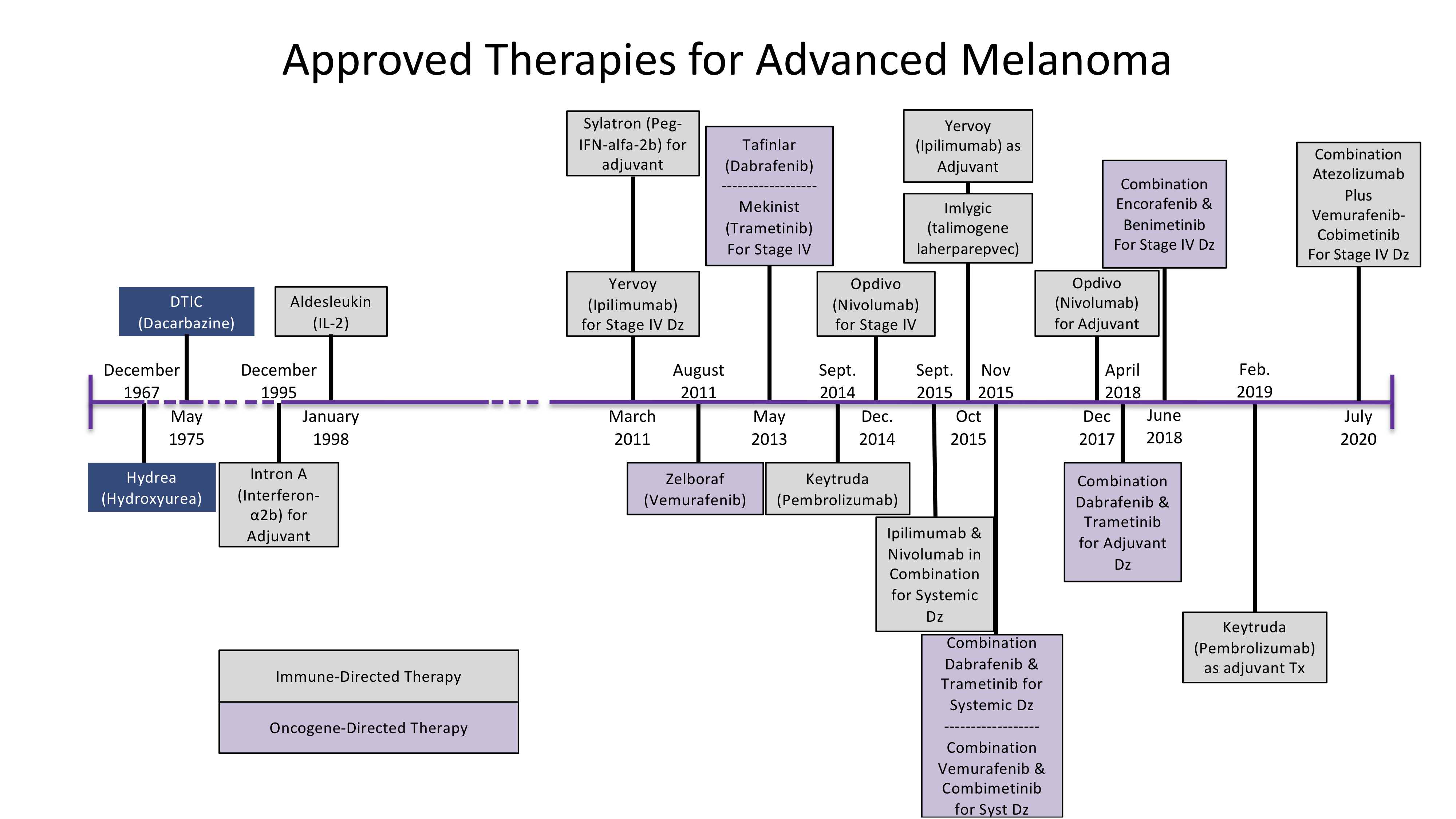
Timeline of FDA-Approved Medications for Melanoma
This site represents our opinions only. See our full Disclaimer and Terms of Use Agreement