Pearls from the Cascade
Overview & Learning Objectives
- The objective of this lecture is to provide a few clinical pearls from Coagulation Cascade
- There is a slide component, which is used for didactic purposes, as well as a monograph section for background (see below)
- In part 1, we will review the clotting cascade and introduce a methodology to approach clinical scenarios where aberations in the PT and PTT are key elements
• The vascular phase
• The platelet phase
• The plasma phase
In this lesson, we will focus on the plasma phase, in which the coagulation factors are activated
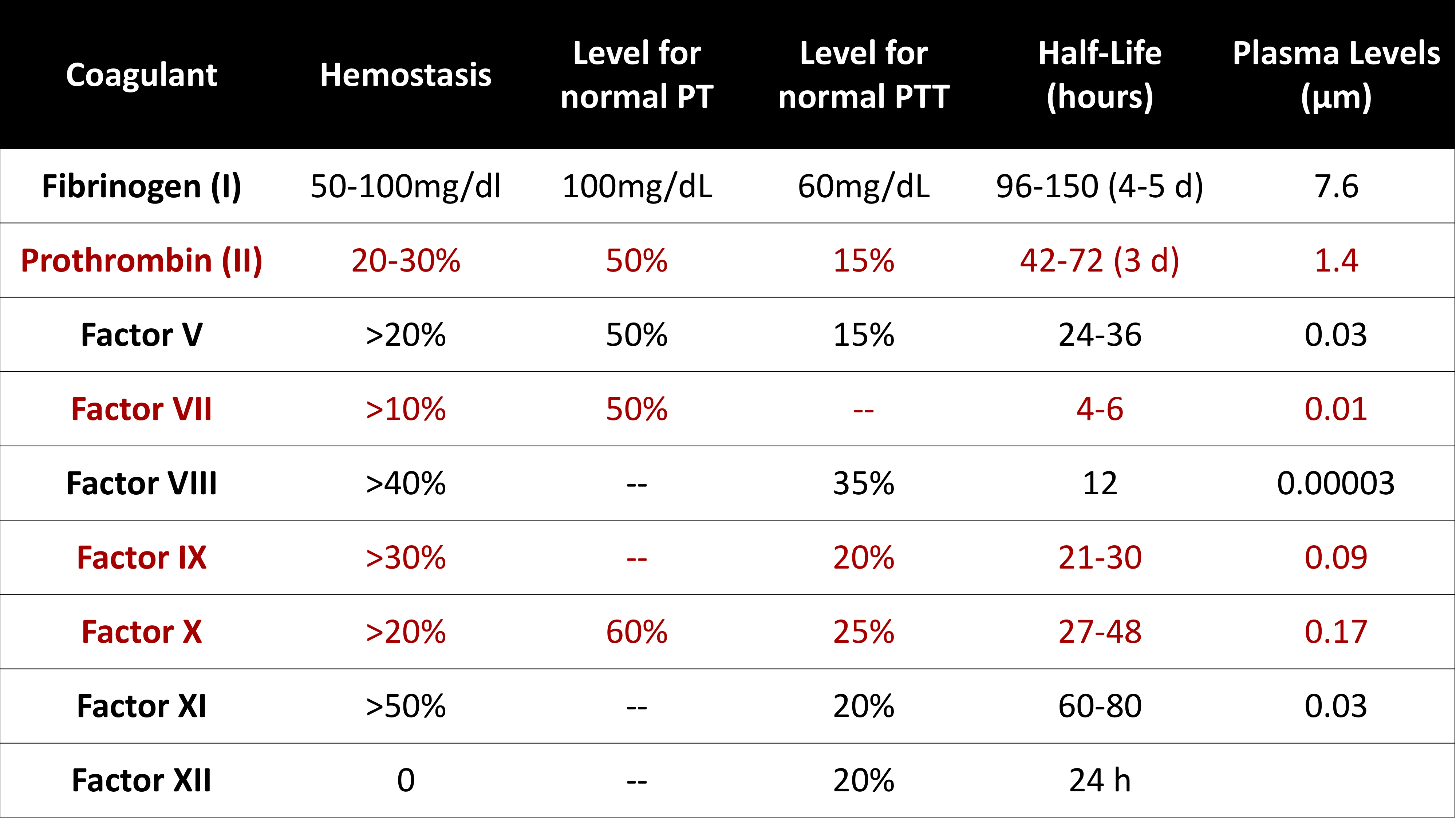
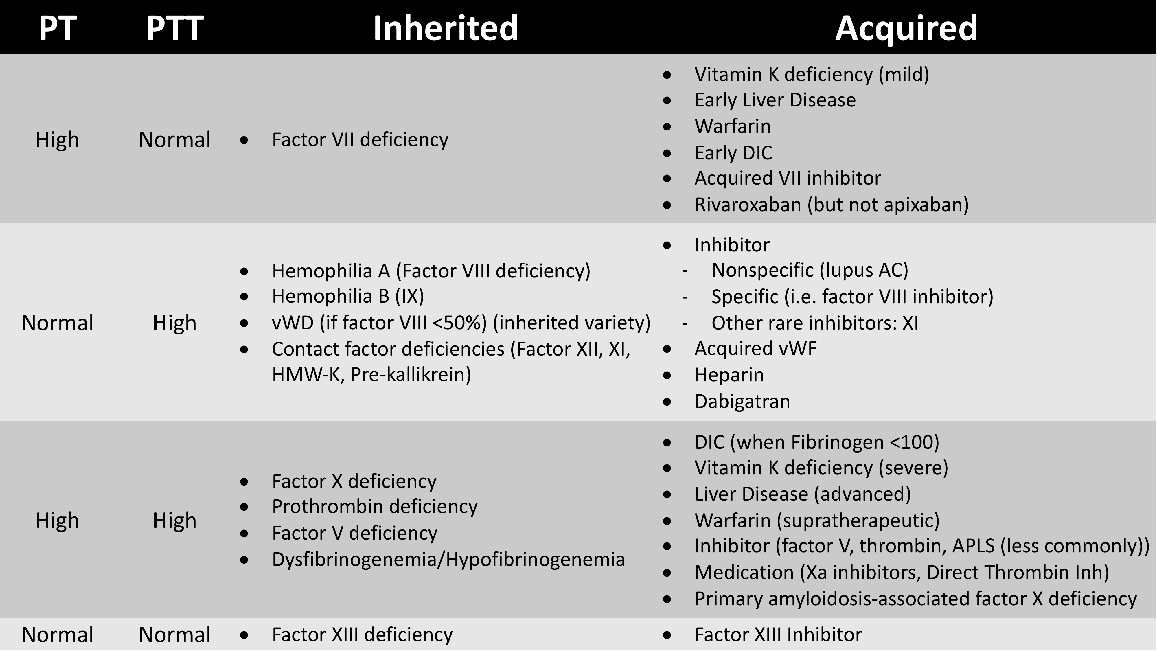
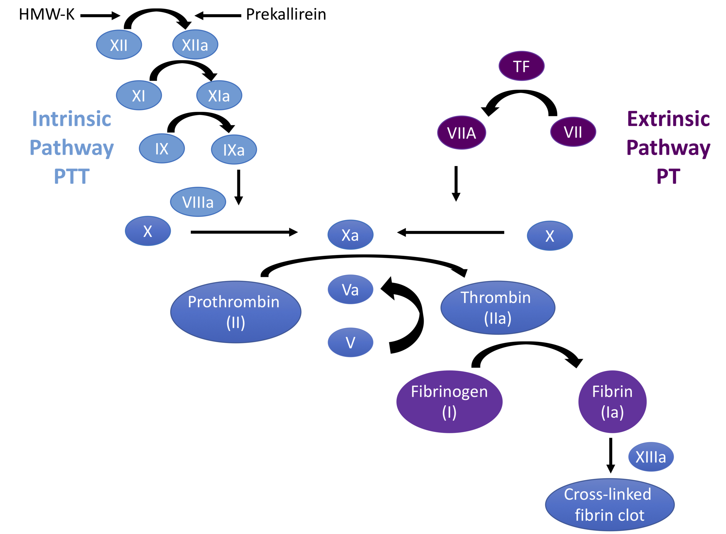
Although the Clotting Cascade is very helpful in evaluating patients with coagulopathies, it is very important to realize that In Vivo coagulation follows a very different set of pathways
We will provide a brief overview of this pathway in the section below
Conceptually, the clotting cascade has been broken down into 3 stages:
1) Initiation
2) Amplification
3) Propagation
Let’s highlight key components of those stages in the following sections
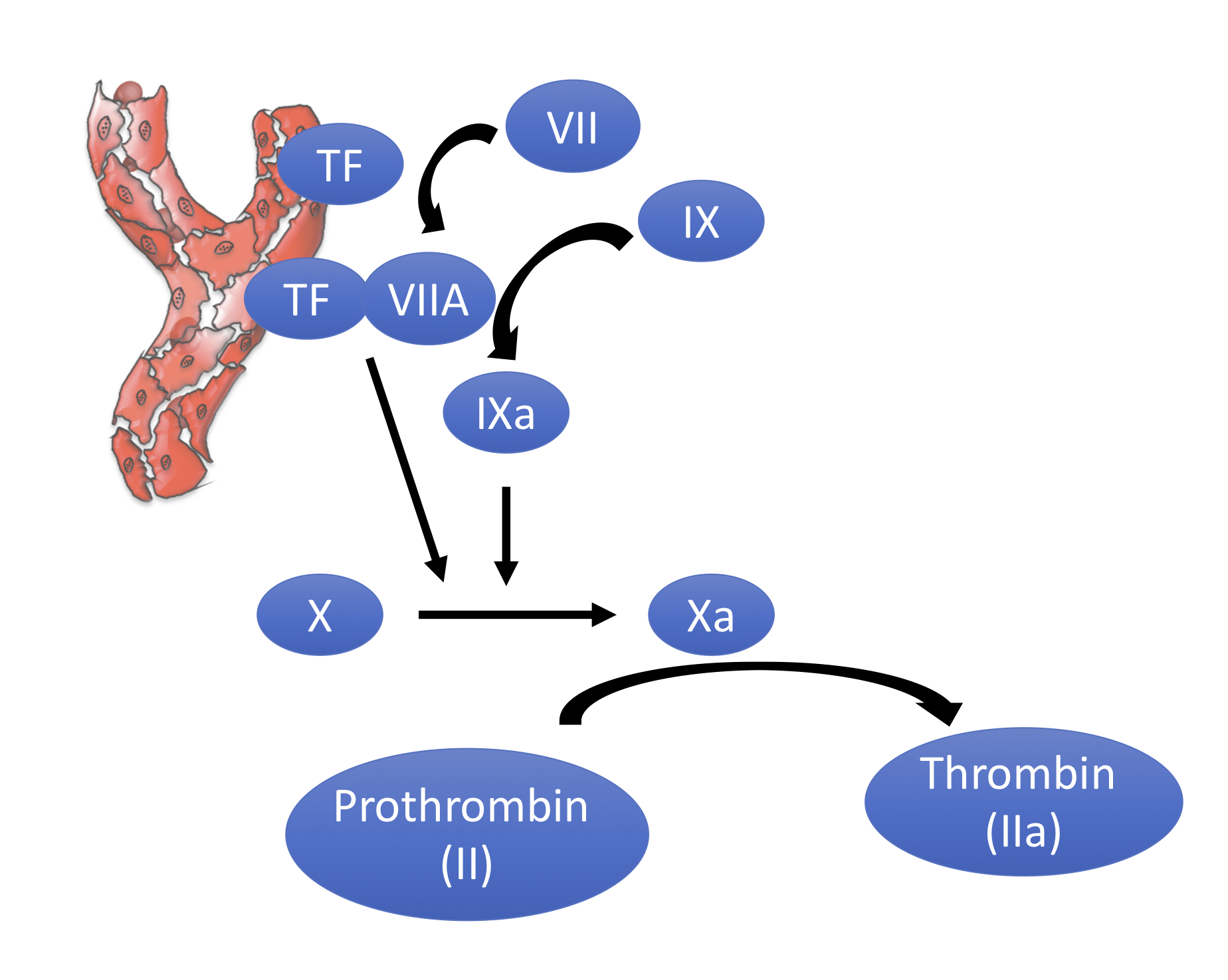
In the body, coagulation is initiated with Tissue Factor is exposed following damage to blood vessels. Following this, TF binds to Factor VIIa. These are the two key entities that make up what is referred to as the Extrinsic Pathway in the clotting cascade.
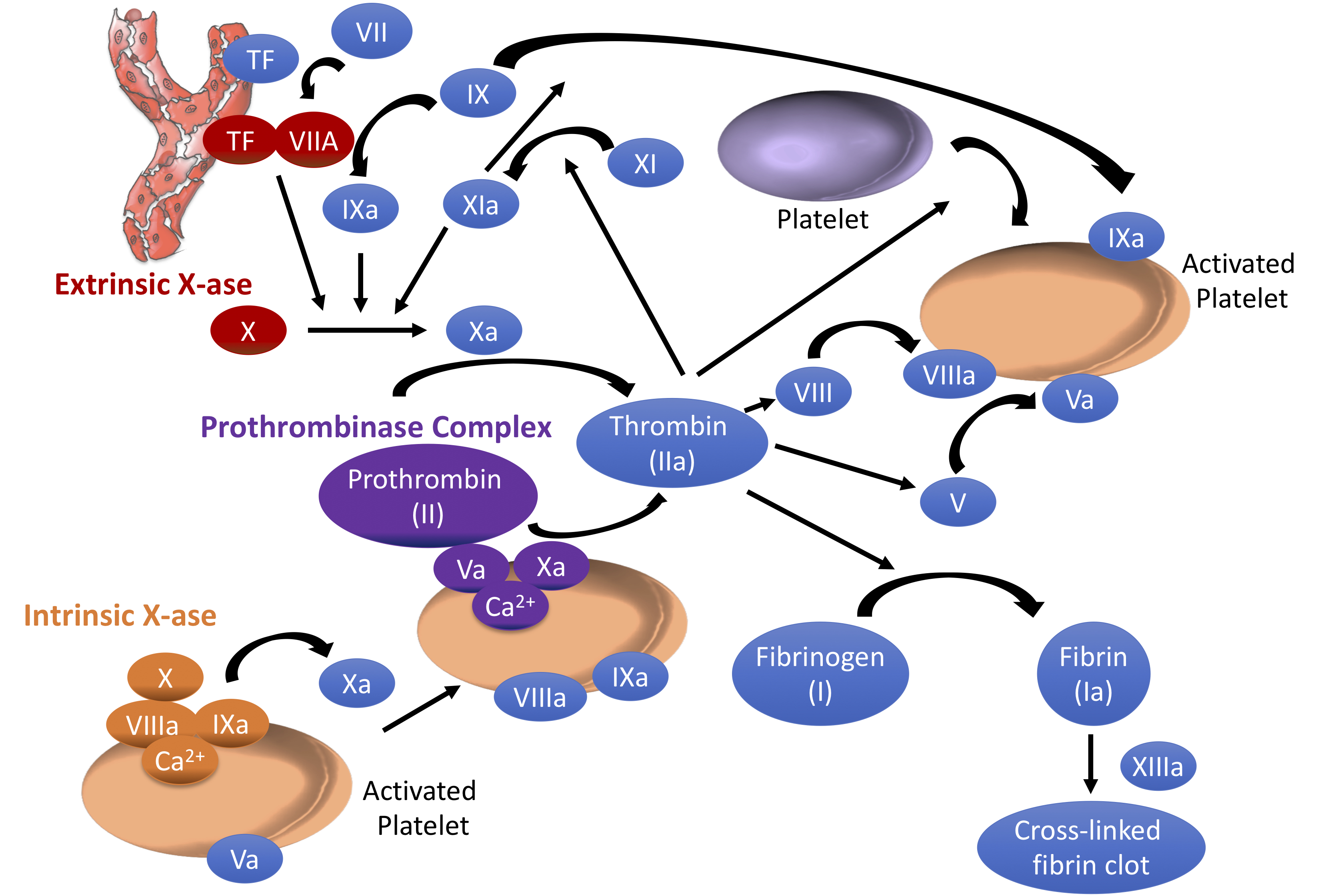
Following initiation of the Extrinsic Pathway, coagulation progresses down what is often referred to as the “Common Pathway”, in which a small amount of Factor Xa is generated, followed by a small amount of Thrombin (see Figure below)
 As seen above,
 Tissue Factor serves as a cofactor for the production of activated factor VII (Factor VIIa). Tissue Factor then binds Factor VIIA. Thus, initiation is driven by the “extrinsic” pathway, as stated above.
The Factor VIIA/Tissue Factor complex that was created then activates Factor IX to IXa
This complex of Factor VIIa as the protease and Tissue Factor as the cofactor and Factor X as the substrate is referred to as the Extrinsic X-ase (see Figure below) 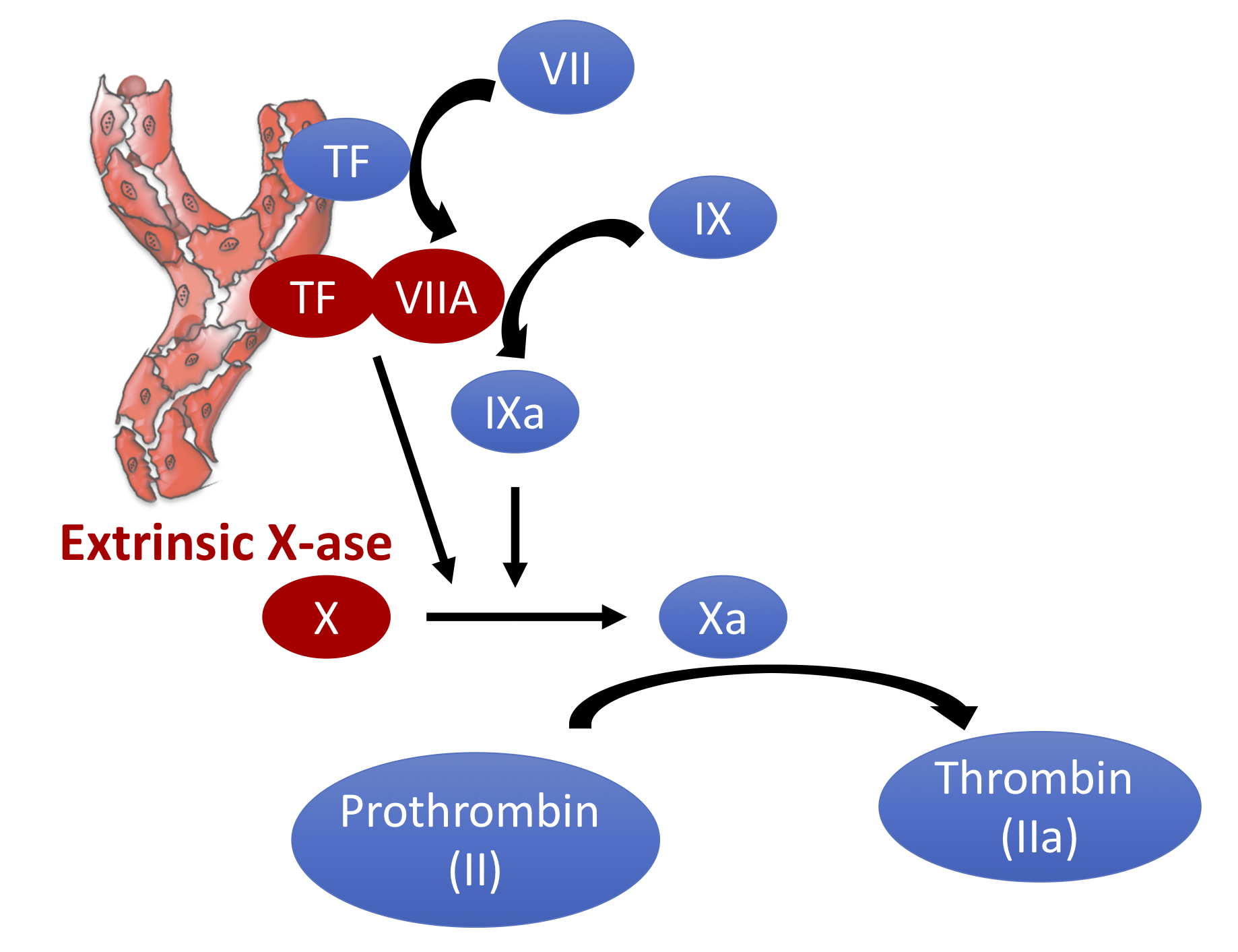
Of note, Factor X is also activated by activated Factor IXa
Activated X (Factor Xa) subsequently produces small amounts of Thrombin by activating Prothrombin
In total, Initiation only produces about 5% of the overall thrombin
The remaining 95% is generated by the `Intrinsic Pathway
There is a mechanism by which after the initial generation of thrombin, negative feedback pathways are activated via TFPI
Xa interacts with tissue factor pathway inhibitor (TFPI) and turns off the extrinsic arm (see figure below) 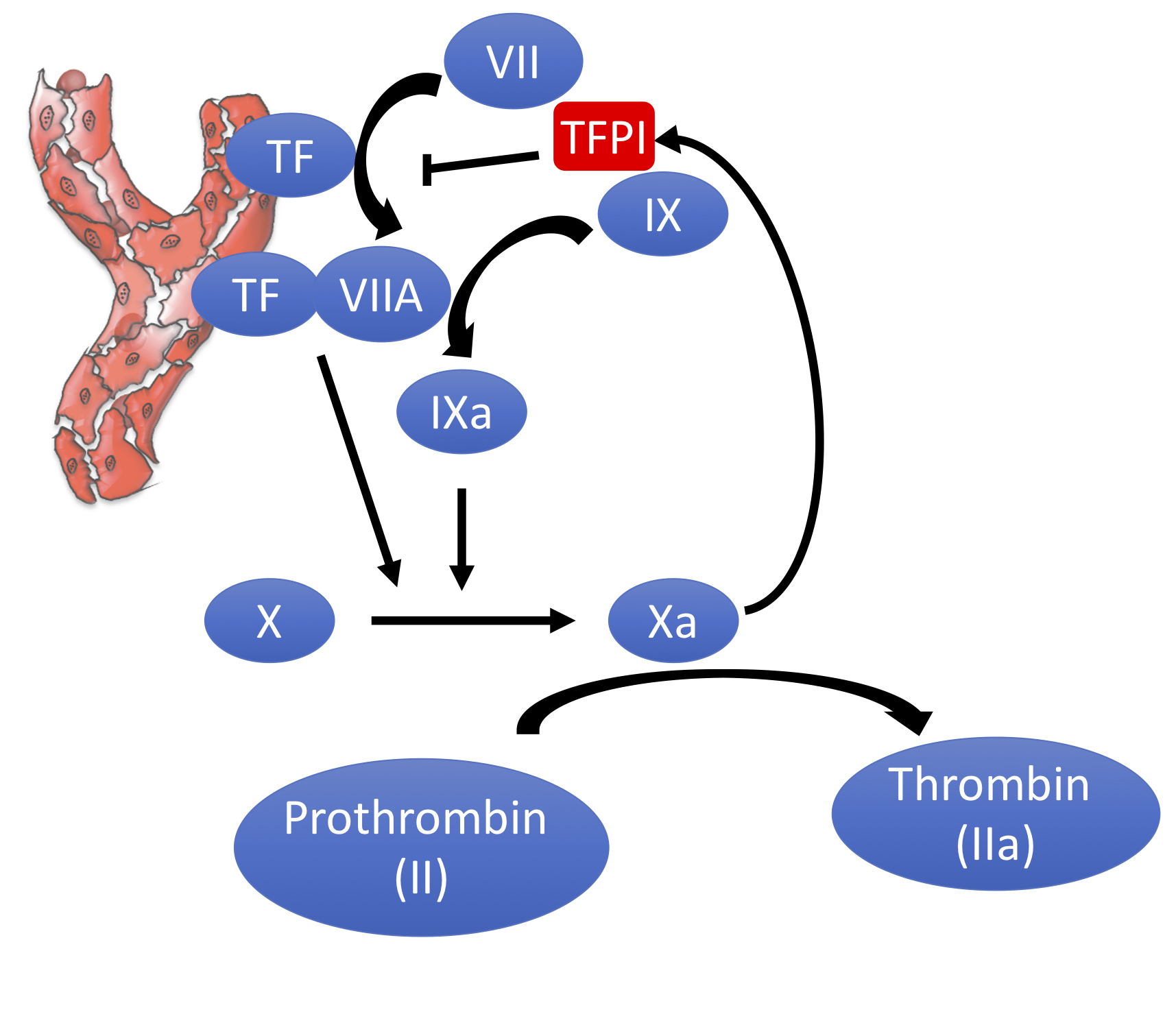
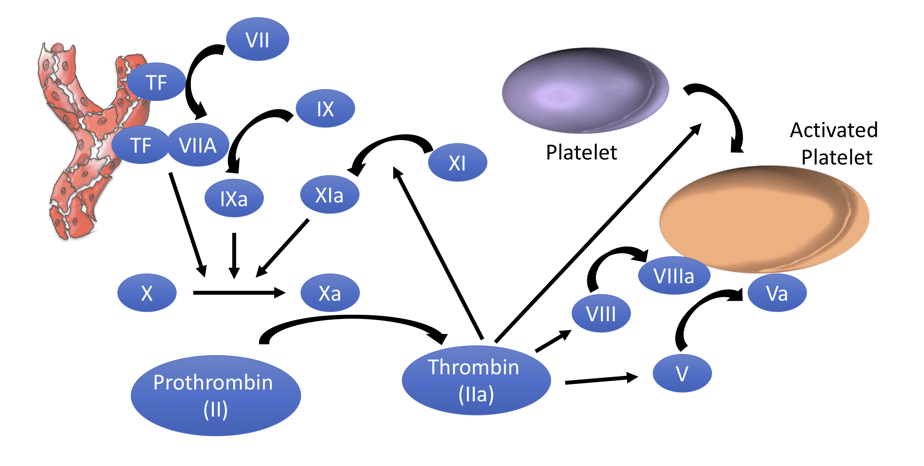
Thrombin plays a pivotal role in propagating the coagulation cascade via several mechanisms:
- Activating
Factor XI(XI -> XIa).XIathen contributes to moreIXa& thus generation ofXa. Consequently, there is more conversion ofProthrombintoThrombin - Activating platelets via PAR receptors (protease-activated receptors). Activation of platelets leads to exposed anionic phospholipids, which support the assembly of the multi-component enzyme complexes. Activating platelets secrete
Factor Vfrom their alpha granules
- Activating
Factor VIII
- Activating
Factor V
(see Figure Below)
 Although challenging to memorize in the abstract, understanding the factors in